Comparison of the calvariums of Homo naledi and Ceprano
By Dr Rob Walsh
Lewis Research Unit
This paper is designed to show the similarities/differences between the newly described H.naledi with the reconstructed Ceprano calvaria discovered in Italy in 1994.
In the case of H. naledi, a composite skull has been made using 4 crania DH1,DH2,DH3 and DH4.
These crania are examined in detail in Lee Berger & co, paper released in September 2015 in eLife sciences publication. In this paper, H. naledi is compared cranially and morphologically with an array of hominin, mainly African.
In this paper I shall solely concentrate on the composite H. naledi’s cranial statistical values with the Ceprano calvaria.
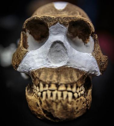
Homo naledi cranial morphological characteristics
The cranium is small at 560 cc. It exhibits frontal bossing, sagittal keeling a post-bregmatic depression, a well-developed supraorbital torus delineated by a distinct supratoral sulcus, with temporal lines on the posterior of the supraorbital torus.
The cranium also has an occipital torus with an external occipital protuberance, but only a slight post-orbital constriction. Also it has a small postglenoid process a slight crista petrosal, and a laterally inflated mastoid process.
The cranium does not have the long and low cranial vault that distinguishes H. erectus nor a metopic keeling. H. naledi has a distinct external occipital protuberance as well as a tuberculum linearum.
A flat and squared nasoalveor clivus and an anteriorly shallow palate.
There is some parasagittal keeling present between bregma and lambda areas. H. naledi has its maximum cranial width in the supramastoid region rather than in the parietal area.
Overall the crania of H. naledi is small and compares better with early Homo (Habilis) than later homo (erectus). (Lee Berger et all 2015)
Ceprano calvaria morphological characteristics
The ceprano has its own cranial morphology. The cranium has had two reconstructions. These characteristics from the first reconstruction include:
The anterior/posterior of the calvarium are quite prominent in their projection with a noticeable fortalis and torus occipitalis. The glabella-inion axis gives the total length
of the calvarium as 208 mm.
A side view of the calvarium shows a roundish aspect with a low frontal vault, seen from the opisthocranion. The inion and the occipital torus has a sulcus supratoralis which is slight in appearance.
The measurement of 115 degree angle shows a sharp bend between the occipital squama and the nuchal plane. The vertex and bregma measure 64 mm.
The front of the calvarium shows an inclination of 50 degrees and the posterior of
the calvarium shows a 51 degree inclination over the occipital squama.
The frontal torus, as well as being low, is rectilinear and has a bulky ridge that
occupies the supraorbital margins and glabella region. The supraorbitals are 20 mm at their maximum height with the height of the glabella midline at 18 mm. Beyond the
frontal torus the calvarium projects flattened sides, and a median tuberosity, without any traces of a metopic suture.
In the posterior region, the temporal line emerges from the torus angularis, without much of a ridge. Near the temporal margin are found slight radial impressions of varying length. These are linked with the original temporal muscles.
The actual temporal bone which remains, is situated in a low position also appearing elongated.
The temporal bone nicely offsets the large mastoid, which in turn has its lateral surface lying below the supramastoid crest level. The mastoid is distinctive as it extends quite far back on the calvarium, and seems to bend inwards slightly.
The incisura mastoidea, is quite wide in relation to its length, and it is noticeable how the supramastoid crest is connected with zigomatics arch's roof, which gives the impression of originally covering the length of the fossa temporalis.
Much was made of the shear depth of the glenoid fossae, with its anterior part infringing on the preglenoid plane, and the posterior only being separated by the
crista post-glenoidal from the external auditory meatuis, and therefore giving the distance for the glenoidal fossae as 152mm.
A 2.5mm distance has been measured from the supramastoid crest and a noticeable swelling in the temporal region.
The calvarium bends inwards towards its middle below the supramastoid crest, before
finally aligning with the mastoid contours.
Unfortunately the fragmented calvarium with slight distortions poses queries over the original point of the maximum lateral prominence on the left side.
At the posterior of the calvarium the low and broad position of the occipitalis torus is
The asterion-breadth is 133mm and the height of the occipital area index is 48.1mm.
The occipitalis torus, jets out, but has a smooth surface, which crosses in a diagonal
or oblique direction, with the left part slightly larger than the right, running at an elevated level. This has been explained as a congenital malformation.
The nuchal area is concave at its posterior, and the endinion-inion has a slant which has the endinion some 22mm below the inion.
The cranial bones are unusually thick, with the right parietal reducing in thickness from 11.5mm (base) to 8mm (vault).
The millet seed method was used to calculate the cranial capacity at 1,185ml. (Ascenzi et all 1996) (R Walsh 2001)
From the second reconstruction by Ron Clarke, the main differences were:
The Ceprano calvarium underwent a second reconstruction to try and incorporate
much of the remaining bone fragments and thereby reduce its incomplete appearance.
Dr Clarke was able to reduce the orbits length by moving the frontal area backwards, and by also rotating the frontal area downwards, in relation to the calvariums posterior. He thereby significantly reduced the gaps in that area.
Dr Clarke located the coronal suture on the calvaria’s anterior margin, which he aligned with the coronal suture of the frontal, once he had moved that particular bone posteriorly.
Both sides of the occipital region were originally cased with plaster. This indeed gave the calvarium a distorted view, (as mentioned in my conclusion on the first reconstruction),
with the asterion - breadth measuring 133mm and occipital area index at 48.1mm .
Prof Clarke removed this plaster, which he found was unnecessary, as the bones fitted together quite well without the enhancement of plaster.
He also systematically pieced together the right sides parietal and temporal bones with the occipital along the lamboid suture.
The left hemisphere of the calvarium enabled Dr Clarke to complete the temporal and posterolateral corner of parietal passing into the occipital at the asterion.
Among the previous undefined bone fragments, Dr Clarke located two pieces of orbital plate. Their correct positioning in the calvaria, assisted in making a correct alignment of the left sphenoid. This was then correctly connected to the left anterior half of the temporal and adjoining parietal.
He also attributed a previously unidentified piece, to the posterior of the right zigomatic arch and to the right temporal bone.
Dr Clarke then removed a fragment from the right sphenoid, and was able to join both front and back of the calvaria, with all areas relating to the calvaria’s anterior and posterior correctly aligned.
Dr Clarke concluded that many of the initial errors in the calvariums first reconstruction previously attributed to congenital malformation or/and post- mortem deformation, were in fact due to errors in the reconstruction.
Other points to mention which are immediately apparent in Dr Clarke’s reconstruction are:
1) The thickness of the temporal at asterion = 22mm (at 19mm only the cranium
Sangiran 31 compares favourably).
2) The maximum length has been reduced from 208mm to 193mm.
3) The angle of the frontal relative to occipital profile, has now been altered. The right aspect shows that the frontal is now rotated clockwise relative to the occipital profile and the braincase is much shorter.
4) Cranial capacity has been reduced from 1185ml to 1057ml (in line with OH9 at 1067cc). This measurement falls within the Rightmire range of Homo erectus crania size between 850cc to 1225cc.
5) Original construction showed the widest point on the braincase across the temporal squama. The new reconstruction changes the point of greatest width to the supramastoids at 161mm.
6) The minimum breadth relates across the frontal at 112mm.
7) No metopic keeling as with OH9.
8) A low and short cranial vault, with a prominent medio - lateral expansion, and its maximum breadth at the level of the crista supramastoidea.
9) (a) Continuous supraorbital torus/bursoidal in aspect.
(b) Calvaria has a continuous supraorbital torus of significant build.
This assists in the calvarias bursoidal shape.
10) (a) There is a noticeable postorbital constriction index;
(M.9/M.44 X 100).
(b) The calvariums frontal trait is not as extended as in other erectus. Its index
of minimum frontal breadth/biorbital breadth is only 84.8. Compare this with
other potential male Homo erectus crania; (i) KNM-ER3733 = 87.5
(ii) Sangiran 17 = 86.1 (iii) Sambung. = 89.5.
As you can see the second reconstruction of the Ceprano calvaria greatly changed its morphology and overall appearance. (Ascenzi et all 2000) (R.Walsh 2001)
Similarities of the two hominin crania
There are some similarities between the two hominin crania.
Both have a Post orbital constriction, sagittal keeling and supraorbital torus. These differ in appearance, but does show the different hominins have similar features.
naledi has a post glenoid process, whereas Ceprano has a pronounced glenoid fossa. Also H.nalidi has an inflated mastoid process and Cerano has supramastoids at 161mm.
Ceprano is generally larger then H. naledi, which could be due to its more recent antiquity at roughly 400 kyr (G.manzi &co). A date for H. naledi has yet to be established.
Conclusions
From comparing the two sets of data derived from H.naledi and Ceprano crania, it appears
That the two are not closely related. H.naledi fits in well morphologically speaking with the smaller homo sp. in Africa.
Lee Berger and co, have attempted to create a new species of hominin in naming this find H. naledi, and this may well be the case. However my comparisons do show that Ceprano is a larger and robust individual, again going on just crania size.
There are similarities in the two crania, but these are superficial and can be attributed to a range of ancient hominin crania. Ceprano is an enigma, as it has been recently re-dated at 300-400 kyr, but exhibits a more ancient morphology comparable with many H. erectus from Africa (OH9) and from China (Zhoukoudian assemblage).
Homo naledi’s affinities as I’ve mentioned, may lay with more primitive habilines from Africa. A date for the H. naledi will give it a better placement on the hominin tree of life.
Of course there may be a relationship between the two in time and space. But this is tentative and the geographical location between the two suggests the contrary.
Acknowledgements
Thanks goes to Professor Lee Berger and team for a detailed description of the H. naledi crania used in this paper for comparison purposes.
Thanks also to the late Professor Antonio Ascenzi for allowing me to originally examine the Ceprano calvarium and describe it.
Bibliography
Ascenzi, A., I. Biddittu, A. G. Segre, P.F. Cassoli, E. Segre-Naldini. A calvarium of Late Homo erectus from Ceprano, Pleistocene, Italy. (Journal of Human Evolution, 31, 409-423, 1996)
Ascenzi, A., F. Mallegni, G. Manzi, A. G. Segre, E. Segre-Naldini, A re-appraisal of the Ceprano calvaria with Homo erectus, after the new reconstruction. (Journal of Human Evolution, 39, 443-450, 2000)
Berger. L.R., J.Hawkes, et co. Homo naledi, a new species of the genus Homo from Dinaledi chamber, South Africa. (elifesciences, September 2015)
Walsh, R., The Ceprano calvaria enigma, (Lewis Research Unit, Independent publication 2001)
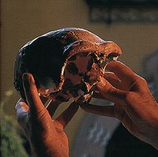
The Ceprano calvaria